
SEE MORE. PROTECT MORE.
Advanced Retinal Imaging
Technology that helps us see more, understand more, and care for your vision with greater detail.
Comprehensive
Retinal Exams at Ironwood Vision Center
Your retina plays an important role in maintaining healthy vision, and regular retinal evaluations help identify changes before they become more serious. At Ironwood Vision Center, we use advanced Eidon® retinal imaging technology to provide a detailed view of the back of the eye during comprehensive eye exams.
Eidon captures a wide-field image of the retina, allowing our team to monitor eye health, compare changes over time, and identify potential concerns with greater detail than traditional imaging methods.

ADVANCED IMAGING TECHNOLOGY
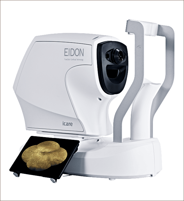
Meet EIDON
EIDON is an advanced retinal imaging system that captures high-resolution, wide-field images of the retina — the light-sensitive tissue at the back of your eye.
The detailed images allow Dr. Garn to examine retinal structures, document your eye health and compare images over time. This can make subtle changes easier to identify and monitor from one visit to the next.
Quick • Comfortable • Noninvasive
WHAT WE'RE LOOKING FOR
A Window Into Your Eye Health
Detailed retinal imaging can help Dr. Garn identify or monitor signs associated with:
Early detection and consistent monitoring can make an important difference in protecting your vision.
WHY EIDON?
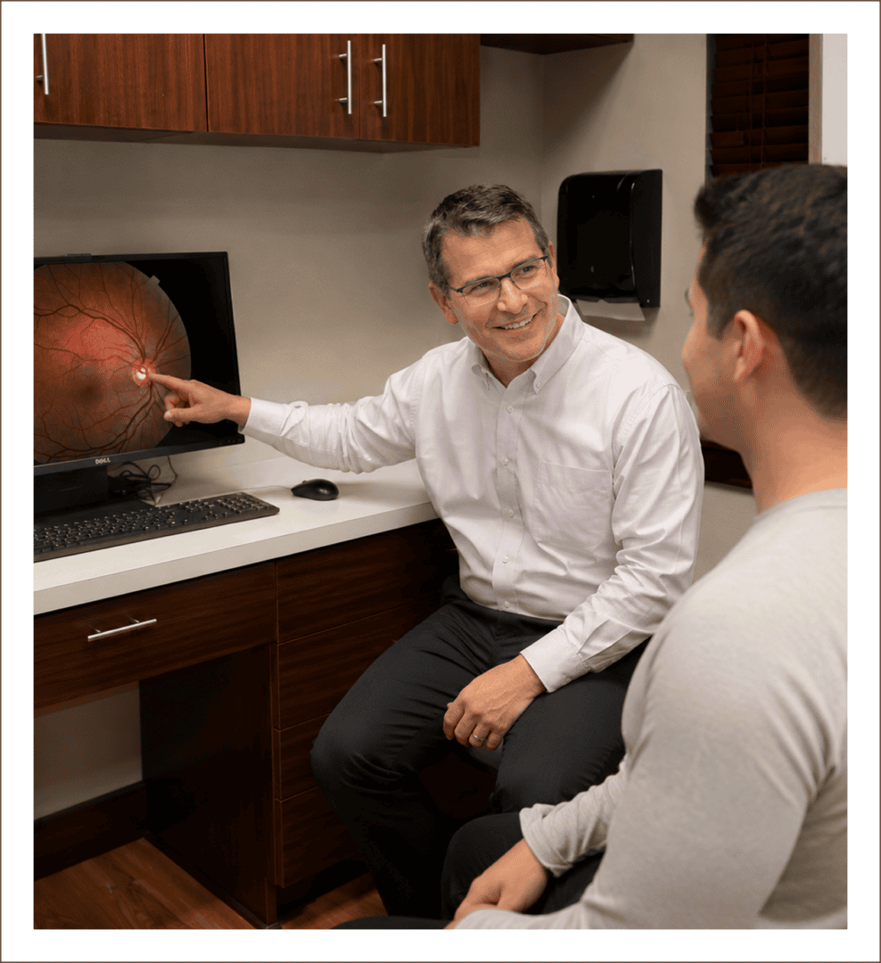
Detailed Imaging. Personalized Care
EIDON gives Dr. Garn a detailed view of the retina that becomes part of your ongoing eye-health record.
ADVANCED RETINAL IMAGING
See Blood Flow in the Eye-Without Dye
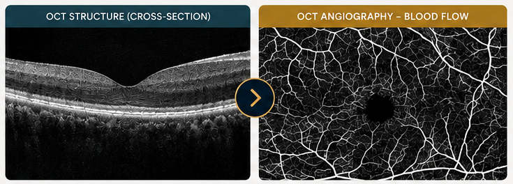
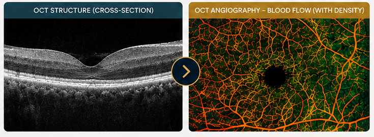
Our Topcon Maestro2 now features OCT Angiography (OCT-A), an advanced, non-invasive imaging technology that allows Dr. Garn to visualize blood flow within the tiny vessels of the retina and choroid.
With the touch of a button, OCT-A captures detailed vascular information without the need for contrast dye injections. It also allows Dr. Garn to examine different layers of the retina individually, providing another level of information when evaluating retinal health.

NO DYE INJECTION
Comfortable, non-invasive imaging.

FAST & NON-INVASIVE
Quick scans for a comfortable experience.
LAYER-BY-LAYER ANALYSIS
Examines vascular information at different retinal depths.

OCTA DENSITY
Quantitative information that helps evaluate vascular changes.

ADVANCED TECHNOLOGY. MORE INFORMATION. PERSONALIZED CARE.
The Maestro2 provides instantaneous vascular flow information together with comprehensive segmentation to enable advanced diagnosis.
OCT Angiography includes OCTA Density.
OCT ANGIOGRAPHY (OCT-A)
Instantaneous vascular flow information without the need for contrast dye injection.
OCT ANGIOGRAPHY (OCT-A)
Instantaneous vascular flow information without the need for contrast dye injection.
OCTA DENSITY
The OCTA Density is defined as the ratio between the high signal area and low signal area and it is displayed in color and/or number.

From detailed structure to detailed blood flow.
OCT Angiography gives Dr. Garn the advanced information needed to evaluate, monitor and manage a wide range of retinal conditions.
ADVANCED IMAGING • DEEPER INSIGHT • PERSONALIZED EYE CARE